The jugular veins are an important part of the drainage system of blood carried from the head and brain to the lungs, where the blood is reoxygenated. The external jugular veins can be seen in the neck alongside the carotid arteries. They carry a great deal of blood away from the head and face, and if injured, tend to bleed a lot.
The External Jugular Veins
The external jugulars are part of three separate jugular veins, all located in the neck. There are essentially three jugular veins that together drain blood from the head and neck into the lungs. These include the following veins:
- The external jugular veins
- The internal jugular veins
- The anterior jugular veins
Without the adequate functioning of these veins, the head would fill with blood and there would be significant edema of the face, scalp and neck. These are very large and very exposed veins in the neck. Any injury to these jugular veins can happen as a result of penetrating trauma which can be life-threatening if not repaired surgically very quickly.
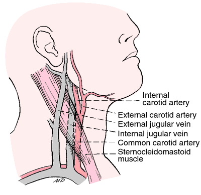
Structure of External Jugular Vein
The external jugular vein is created by the confluence of two separate veins: 1) the posterior auricular vein which is the vein responsible for draining the top of the head and the area behind the outer ear, and 2) the retromandibular vein, also known as the posterior branch of the external jugular, which is important in the drainage of deoxygenated blood from the face.
The two veins join together at the angle of the jawbone, also called the mandible. The vein continues on to enter the thorax beneath the clavicle, finally terminating in the subclavian vein that goes on toward the heart, where blood is pumped into the lungs. In the lungs, the blood is given the oxygen it needs to supply the body with fresh blood.
There are several, minor tributary veins that also enter the external jugulars. In particular, the posterior jugular vein, the suprascapular veins, and the transverse cervical veins are minor tributary veins that drain into the external jugulars in the area of the neck.
Why It Is Important
Because of the superficial location of the external jugular vein, it is prone to being injured from a knife wound or other penetrating injury. It isn’t very deep to the skin so that a minor slash with a knife can easily puncture the vein, leading to excessive bleeding. In some cases, air from the outside can enter through a slash in the jugular vein and can get lodged in the right atrium of the heart.
The external jugulars have at least two separate pairs of venous valves that don’t have much function. They don’t prevent blood from regurgitating back up to the head and they don’t prevent blood flow from the face from travelling down the neck. Their exact purpose is unknown.
If you search for external jugular vein for getting to know the external jugular vein IV, please watch the video below:
More Kinds of Jugular Veins in the Neck
As mentioned, the external jugulars are part of a system of jugular veins, all of which converge in the neck area to eventually enter the right atrium of the heart.
1. Posterior External Jugular
It receives blood from the occiput of the head, which is the area of the head in the back. This includes the scalp and internal structures of the back of the head. The posterior external jugular collects blood from the occiput and sends it around the side of the neck to join with the external jugular vein.
2. Anterior Jugular Veins
These begin in the area of the hyoid bone, which is located in the front of the neck. It travels downward in front of the sternocleidomastoid muscle, which is a major muscle that is involved in the turning of the neck. At the bottom part of the neck, it goes beneath the sternocleidomastoid muscle, where it dumps blood into the external jugulars. In some cases, it doesn’t drain into the external jugular at all but ends up directly into the subclavian vein in order to travel down into the thorax where the superior vena cava enters the right atrium.
3. Internal Jugular Vein
This is the vein that drains blood directly from the brain as well as parts of the face and neck. The internal jugular vein connects with the transverse sinus at the jugular foramen, which is the area at the base of the human skull that is the opening for the blood to drain from the veins of the brain to the outside of the skull itself.
Veins themselves aren’t as particular about their location and access points as that about the arteries, so there may be some variations in the appearance and the exact location of the vein. For example, the external jugulars might not be a single vein but may descend the outer portion of the neck as double set of "twin veins" that both lead to the subclavian vein in the lower part of the neck and the upper part of the thorax. This doesn’t affect circulation at all and is a normal variant to the single vein that most people have.