Have you ever seen someone with a red, droopy eye that almost appeared as if it were sinking into the eye socket? Then you may have encountered someone with Horner’s syndrome. Also known asHorner-Bernard syndrome or, more formally as oculosympathetic palsy, this condition always results from some other medical problem.
Horner’s syndrome is a relatively rare medical condition that comes as a result of another medical issue. It can strike anyone of any age, though it happens to men more frequently than women or children. Studies show that 5 out of 6 victims of Horners syndrome are male. So what causes this syndrome and what can be done about it?
Symptoms of Horner’s Syndrome
Some of the symptoms displayed by a person with Horner syndrome include:
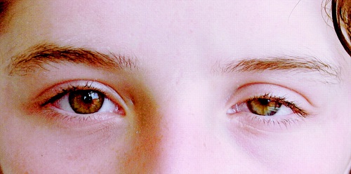
- One pupil being smaller than the other, a condition known as miosis.
- There is also ptosis, which is medical terminology for a drooping eyelid.
- The eyeball may appear to be sinking into the orbital cavity and may be bloodshot. The lower eyelid could be swollen.
- The affected side of the face may not sweat or flush the way the other side does.
- The affected eye may experience reduced tear production and in rare cases, it can cause the irises of the eye to appear to be different colors, especially in children less than a year old.
What Causes Horner’s Syndrome?
Horner’s syndrome is a collection of symptoms that affect the eye and face. It’s caused by the disruption of the path used by nerves to travel from the part of the brain known as the hypothalamus to the eye and face on one side of the body. The hypothalamus is in charge of blood pressure, heart rate, pupil size, and other functions that help your body adjust quickly to changes in the environment.
There are three types of nerve cells, or neurons, in the nerve pathway that are affected by Horner’s syndrome. One type is called first-order neurons. This path travels from the hypothalamus through the brain stem then spreads into the upper spine. The nerve function in this area can be affected by events such as:
- Neck trauma
- Tumor
- Stroke
- Cysts in the spinal column
Second-order neurons travel away from the spinal column, through the upper portion of the chest and continue on into the side of the neck. Events that can result in nerve damage in this location may include:
- Traumatic injury
- Lung cancer
- A damaged aorta, the main blood vessel that leads away from the heart
- Tumor in the protective coating that covers nerve endings (myelin sheath)
- Chest cavity surgery
Third-order neurons run through the side of the neck and into the skin on the face and to muscles that regulate the eyelids and irises. Nerves in this region may be damaged by:
- Migraine
- Cluster headache
- Carotid artery damage
- Jugular vein damage
- Infection or tumor near the skull’s base
When the condition affects children, there may be other causes such as:
- A congenital birth defect in the aorta
- A neuroblastoma (tumor found in the nervous or hormonal systems)
- Trauma to the shoulders or neck during birth
Sometimes it is impossible to tell exactly what is causing Horner’s syndrome. It is then called idiopathic Horner syndrome.
Treatment for Horner’s Syndrome
If you or someone you care about is showing symptoms of Horner’s syndrome, you should seek medical advice. Remember, Horner’s syndrome is always the sign that there is an underlying medical problem. If the symptoms appear suddenly or following a traumatic injury, treat it as an emergency and go to the nearest emergency room or call 911.
There is no treatment specifically for Horner’s syndrome. Instead, it is necessary to find the condition that is disrupting the nerves’ path. Your medical provider will perform a physical exam and ask you questions like:
- Have you suffered any recent injury or trauma?
- When did the symptoms start?
- Do you suffer from cluster headaches or migraines?
- Have you ever had cancer?
- Do you have pain in your neck, arm, shoulder, or head?
Your doctor may also order imaging tests to help pinpoint the abnormality that is interrupting the nerve’s path. Those might include tests such asMRIs to create detailed images, CTs and X-ray imaging. In case of a child, the physician may also want urinalysis or blood tests conducted that may help detect a neuroblastoma.
Once the problem has been diagnosed and treated, if the treatment is successful, the symptoms of Horner syndrome should be eliminated.